Amnis® ImageStream®X Mk II Imaging Flow Cytometer

The revolutionary ImageStream®X Mark II Imaging Flow Cytometer combines the speed, sensitivity, phenotyping abilities, and quantitative power of fluorescence activated cell sorting flow cytometry with the detailed imagery and functional insights of fluorescence microscopy. This unique combination enables a broad range of applications that would be impossible using either technique alone.
The ImageStream®X Mark II produces multiple high resolution images of every cell directly in flow, including brightfield, darkfield (SSC) and up to 10 fluorescent markers with sensitivity exceeding conventional flow cytometers.
- 40X magnification (60um field of view; 0.5um per pixel; 4um eff depth of field; 0.75 NA). Cells or objects with a diameter less than 60 um are well suited for the ImagestreamX Mark II.
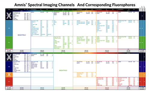
- 5 lasers:
- 405 nm (150mW)
- 488 nm (200mW)
- 561 nm (200mW)
- 642 nm (150mW)
- 785 nm (70mW/SSC)
- 12 total channels: side scatter, brightfield (2) and up to 9 fluorescent colours
AutoSampler for Multiwell Plates
The AutoSampler option for the ImageStreamX Mk II Imaging Flow Cytometer enhances productivity with unattended sample loading from multiwell plates. The fully integrated AutoSampler option allows you to easily perform dose response and time course studies even in primary samples, due to the system’s capacity for imaging large numbers of cells from each sample.
Features
- Ideal for high throughput analysis of suspended cells
- Easy to use acquisition templates allow for multiple experiments per plate
- Unattended operation and in-line bubble detection enables robust operation
- Automatic process logging and error notification
- Automatic sipper rinse between samples and <0.5% carryover
- Automatic sample resuspension via aspiration
- Advanced template function allows groups of wells to be defined for flexible plate configuration
- Automated data analysis
Features of the ImageStreamX Mk II System:
Fast: Analyzes Thousands of Cells Per Second
The ImageStreamX Mk II Imaging Flow Cytometer can analyze up to 5,000 cells/sec with real-time intensity compensation — ideal for rare cell analysis.
Easy-to-Use: Simple User Interface
The intuitive user interface provides real-time plotting and graphical gating, as well as images of every cell. The easy-to-use compensation wizard quickly guides you through setup of multi-color compensation matrices.
Flexible: Fits Your Sample, Your Targets, Your Fluorophores
The ImageStreamX II Imaging Flow Cytometer accepts up to 6 lasers and works with sample volumes of 20-200 µL for added experimental flexibility — perfect for multi-user laboratories.
Efficient: Less Sample Volume, Less Sample Waste
Fluidics that provide up to 95% sample utilization when every cell is required. Recover unused sample for further analysis when orthogonal methods are required.
Adaptable: Any Upgrade, Any Time
The modular design of the ImageStreamX Mk II Imaging Flow Cytometer allows the system to grow with your research needs and budget through a broad range of field-installable options. Most options, including additional lasers, can be installed at the factory or upgraded in the field by Luminex-certified field service personnel.